


Interactive CT & MRI Anat.Lite

Descrizione di Interactive CT & MRI Anat.Lite
★Lite version★
This is the free Lite version of "Interactive CT and MRI Anatomy".
The function is restricted.
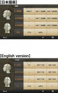
You can only see the transverse CT images of the head.
Please check the operation before purchasing the full version.
★ Details ★
This application is developed for medical students, interns, residents, doctors, nurses, and radiology technicians to understand the essential anatomical terms of the body.
You can learn anatomy by answering the terms by step-to-step questions using a total of 241 CT and MRI images.
A total of 17 images of 3D-CT, MRA and plain X-ray film(particularly the extremities) are included as references.
Other reference images include coronary artery segments defined by the American Heart Association(AHA), pulmonary segments, and liver segments(according to Couinaud classification).
You can enlarge all the images by simple manipulation.
★ Major functions ★
There are 4 major functions.
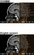
-1) Anatomical mode
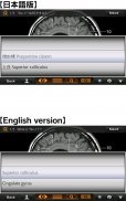
Anatomical terms are overlaid on the images.
It can be used as the anatomical atlas.
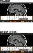
-2) Quiz mode type 1
You select the part of the image by using anatomical term.
Questions will basically appear randomly.
-3) Quiz mode type 2
You select the anatomical term by the part of the image.
Questions will basically appear randomly.
-4) Index
You can find the specific images by using anatomical terms.
★ Intended users ★
-Medical students
-Interns and residents
-Doctrors
-Nurses
-Radiology technicians
-All those who are intrested in CT and MRI anatomy
★ Images(a total of 258 images) ★
Images basically include horizontal, coronal, and sagital planes.
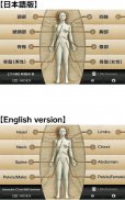
-Head(36 images including CTA and 3D-CT)
-Neck(24 images)
-Spine(19 images including plain X-ray films)
-Chest(61 images including 3D-CT images)
-Abdomen (37 images)
-Pelves: male (9 images)
-Pelvis: female (11 images)
-Extremities (shoulder, hand, elbow, hip joint, knee, foot) (61 images including plain X-ray films)
Editors
Toshiaki Nitori, M.D. (Professor of Radiology, Kyorin University, School of Medicine)
Yasuo Sasaki, M.D. (Manager of diagnostic radiology, Iwate Prefectural Central Hospital)
</div> <div jsname="WJz9Hc" style="display:none">★ versione Lite ★
Questa è la versione Lite gratuita di "Interactive TC e RM Anatomy".
La funzione è limitata.
È possibile visualizzare solo le immagini trasversali TC del capo.
Si prega di controllare il funzionamento prima di acquistare la versione completa.
★ ★ Dettagli
Questa applicazione è stata sviluppata per medici studenti, stagisti, residenti, medici, infermieri e tecnici di radiologia per comprendere i termini anatomici essenziali del corpo.
Si può imparare l'anatomia rispondendo ai termini di passo-a-passo domande utilizzando un totale di 241 TC e RM immagini.
Un totale di 17 immagini di 3D-CT, MRA e film di pianura a raggi X (in particolare alle estremità) sono inclusi come riferimenti.
Altre immagini di riferimento sono segmenti coronarica definiti dalla American Heart Association (AHA), segmenti polmonari, e segmenti epatici (secondo la classificazione Couinaud).
È possibile ingrandire le immagini con una semplice manipolazione.
★ ★ Funzioni principali
Ci sono 4 funzioni principali.
-1) Modalità anatomico
Termini anatomici sono sovrapposte le immagini.
Può essere utilizzato come atlante anatomico.
-2) Quiz tipo di modalità 1
Si seleziona la parte dell'immagine utilizzando anatomica termine.
Le domande saranno fondamentalmente appaiono in modo casuale.
-3) Quiz modalità di tipo 2
Si seleziona il termine anatomico dalla parte dell'immagine.
Le domande saranno fondamentalmente appaiono in modo casuale.
-4) Indice
Potete trovare le immagini specifiche utilizzando termini anatomici.
★ ★ Gruppo target
Studenti -Medical
-Interns E residenti
-Doctrors
-Nurses
La radiologia tecnici
-Tutti Coloro che sono intrested in TC e RM anatomia
★ Immagini (un totale di 258 immagini) ★
Immagini fondamentalmente comprendono piani orizzontali, coronali, sagittali e.
-head (36 immagini tra cui CTA e 3D-CT)
Scollo (24 immagini)
-Spine (19 immagini tra cui pellicole radiografiche plain)
-Cinghiaggio (61 immagini tra cui le immagini 3D-CT)
-Abdomen (37 immagini)
-Pelves: Maschio (9 immagini)
-Pelvis: Donna (11 immagini)
-Extremities (Spalla, mano, gomito, anca, ginocchio, piede) (61 immagini tra cui pellicole radiografiche plain)
Editors
Toshiaki Nitori, MD (Professore di Radiologia, Kyorin Università, Facoltà di Medicina)
Yasuo Sasaki, MD (Direttore di radiologia diagnostica, Iwate Prefectural Hospital Central)</div> <div class="show-more-end">


